www.fluorescencemicroscopy.it
Main menu:
- Home Page
- Microscopes
- Fluorescence
- Description
- Diascopy
- Epi-Fluoresc.
- Illumination
- Installing and Aligning a Mercury Lamp
- Fluorocromes
- Tables of Fluorocromes
- The Fading
- Wavelengths
- Properties
- % of transmission
- Types of filters
- Overlapping Spectra
- Intensity
- Image acquisition
- The Filter's set
- The Filters
- Fluorochrome's praparation
- Sample's preparations
- Fluorescence Sample
- Cytochemical Fluorescence
- Intrinsic Fluorescence
- Gallery
- Phase Contrast
- Polarization
- Darkfield
- DIC
- COL
- Rheinberg
- Brightfield
Illumination
Fluorescence
Illumination in Fluorescence
In a fluorescence microscope can be used, for lighting, a variety of sources, from classic mercury lamp (HBO) or Xenon, to the latest Metal Halide, until the newest: the LED.
This section covers all of these solutions by providing for each of the advantages and disadvantages together with the characteristics of the product.
In general we can say that to generate a excitation light a sufficiently intense to produce an emission suitable to observation, with this technique, it's need to use of compact and powerful light sources. Since the beginning of fluorescence microscopy were used, for this purpose, the discharge lamps. This type of lamps, with power ranging from 50 to 200 Watt (HBO), and 75 to 150 Watt (XENO) are generally powered by a generator, which provides some firing output of gas and allowing the vaporization by burn the vapors resulting in a very high luminous efficiency. The power supply can be provided with a timer that monitors the number of hours of the burner. This is because this type of lamp after a certain period of use lose efficiency and must be replaced, otherwise there is possibility of explosion if used in excess of their estimated lifetime. HBO lamps do not provide, however, a constant light output from the infrared light spectrum to ultraviolet. In fact, most of the emission occurs in the near ultraviolet. The peaks of intensity are located to 313, 334, 365, 405, 436, 546, and 577/9 nm. For other wavelengths in the visible region, the intensity is not as strong but it is still used in many applications of fluorescence. So we can say that in considering the efficiency of a light source, the lamp power is only one factor to consider.
Primary importance is the distribution of intensity at different wavelength, the geometry of the emission from the arc and the opening angle.
Wavelength characteristics
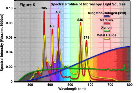
One of the most important variables to consider when choosing a light source for microscopy is the spectral distribution or the profile of the wavelength emitted by the source. Although the characteristic of artificial light sources and some arc lamps is to produce white light that has enough brightness and uniformity across the visible region of the wavelength, the same, for example, does not apply to LED, laser, and some arc lamps (mercury and metal halide). Historically, the fluorochromes were selected for use in fluorescence microscopy because they are specifically excited by lines of greater intensity produced by the mercury arc lamps. This strategy not only served to increase the brightness, but also to take advantage of limited bandwidth in the design of effective two-color mirrors and thin films required for interference filters. In addition, the objectives of the microscopes on the market were often intended to produce a good color correction at these wavelengths.
Even Xenon lamps have been and are still widely used in Fluorescienza, these, in contrast, have no peaks in the spectrum that can be found in mercury lamps, but are perhaps even more useful because their continuous spectrum can be effectively used to excite multiple fluorochromes simultaneously.
In contrast, the emission lines of LEDs are rarely aligned with those of the HBO lamp, then, to use these light sources in fluorescence requires a new assessment of suitable fluorochromes and the corresponding set of filters on the market today. The big optical microscopy houses are still evaluating, and in some cases finalizing, the opportunity to replace the old type of arc lamps with this new technology.
The spectral profiles of different sources of light common to optical microscopy are presented in the figure above. The distinct peaks present in the spectra of the mercury lamp and metal halide occur at 365, 405, 436, 546, and 579 nanometers, some regions of the spectrum are very low as the one between 450nm and 530nm, the excitation band at these lengths is problematic.
On the contrary, instead, the halogen lamp shows a broad spectral profile that has relatively low bandwidth in the ultraviolet wavelengths, but increases gradually before leveling off in the near infrared region. The relative power of the lamp is, however, about 25 percent of the mercury lamp in the center of the visible region (550 nm: the spectral profile of the halogen lamp is shown at 10x as actual output), this makes it attractive for used for certain types of fluorochromes (FITC). Unlike the mercury lamp, xenon lamp has a low but continuous energy profile in the visible region with most of it concentrated at wavelengths of 800 nanometers, this is one reason why in some cases, such as fluorescence using multiband filters, xenon lamps may be the best choice. The metal halide lamps arc (Metal Halide) as spectral lines are functionally identical to those HBO, but produce more continuous energy levels between the lines though with peaks lower, thus making them more useful when used with all the fluorochromes that does not require special peaks
Optical and physical properties of the illumination sources
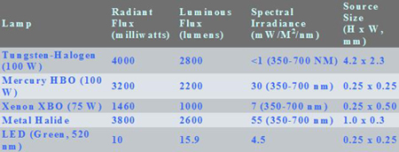
Table 1.

Table 2.
The tables above describes the comparison between the optical and physical properties of the most common sources of illumination for fluorescence optical microscopy. The mercury lamp HBO 100 watt has the highest brightness (luminance average) of the lamps at any level of power commonly used in microscopy, mainly because of very small size of the source (arc). For the microscopist, the spectral content of the light source (in the table above as spectral irradiance) is a very important consideration when comparing different sources of light. The radiant flux defines emission of light at all wavelengths and does not provide information on its spectral distribution (in fact, the number and intensity of various wavelengths actually emitted). This is particularly evident when the photometric units, such as average luminance, are used for the comparison of different sources. Since the photometric units are weighted according to the limited spectral sensitivity of the human eye, the band in the ultraviolet and infrared has an importance factor very small compared to that of green light (at center of the response curve of the human eye ). The comparison between the radiant flux or luminous monochromatic and polychromatic light sources (such as lasers and LEDs) are not significant, if only a limited portion of the spectrum can be used by these sources.
The light sources based on plasma discharge (arc lamps), incandescent (halogen), or stimulated emission in a gas (gas lasers) require a considerable period of time after switching to a balanced position heat, a factor that may affect activities that have in time, space and stability, important elements of success. All lamps that produce a significant level of heat, including light-emitting diodes, also has a dependency on the production of emissions by the source temperature. In many cases, a maximum of one hour is needed until the light source is sufficiently stable to allow reproducible measurements or record video sequences in time-lapse, without significant temporal variations in intensity. Once the proper operating temperature is reached, the halogen lamp is the most stable source of light in conventional time periods of a few milliseconds due to the thermal inertia of the tungsten filament. The LED (light-emitting diode) are able to react, however, very quickly (within a few microseconds), but, when used at maximum power, can also generate a significant amount of heat during the warm-up and, because of their high speed, are affected by power supply instability at high frequencies. In general, we can say that the arc discharge lamps are the most unstable light sources currently used in optical microscopy. Besides the fact that the flicker of the arc deteriorates with age, the light output can also be influenced by electromagnetic fields from power supply or unstable environment.
The new technologies : LED
The compact LED is a semiconductor that emits incoherent light in a narrow spectrum when it is powered. The color of light emitted depends on the composition and condition of the semiconducting material used, and can be near ultraviolet, visible or infrared. LED technology has evolved considerably in recent years, when the first commercial LEDs were introduced in 1968 were able to provide only 0.001 lumens of red light, a level of brightness suitable only for use as indicators. Over the past four decades, LED technology and advanced at a rapid pace, comparable to the rate of advancement of microprocessors.
Now that the LEDs provide sufficient intensity at specific wavelengths required, fluorescence microscopy is able to exploit the full benefits, including: compact size, low power consumption, minimal heat output, fast switching, very stable high emission, long service life. The only drawback: the limited power. To date, in fact, the LEDs with higher power output, 5W, does not yet provide a light output equal to that of an HBO 50W or Xenon 75W lamp. Of course we talk about individual LED and non "chip", or arrays of LEDs, which have a light output so much higher but in a larger amount of space and therefore not suitable for illumination microscopy.
LED technology offers the possibility to choose the excitation wavelength most appropriate for each fluorochrome used. In practice, using this light source we can do without the excitation filter even if it is not always possible or preferable to implement this kind of choice.
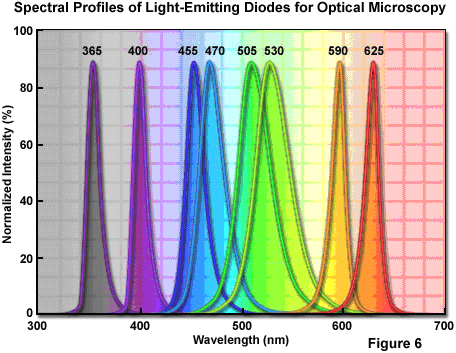
Currently, there are a number of wavelengths ranging from ultraviolet (365 nanometers) to infrared (above 800 nm, see figure above). The only significant difference in terms of fluorescence microscopy is the green-yellow region between 530 and 580 nanometers, but the LEDs that emit at this critical wavelength should be available soon. Note that tests with white LED (cold light) showed a spectral response similar to that of a Blue LED. The spectral width (width at half maximum, FWHM) of a typical quasi-monochromatic LED ranges from 20 to 40 nanometers , which is similar to the bandwidth of excitation of many fluorochromes. Compared to laser light, the wider the bandwidth of the LED is more useful for excitation of fluorochromes. Moreover, compared to the continuous spectrum of arc lamps, LEDs are cooler, smaller, cheaper and provide a mechanism to select multiple wavelengths with fast switching. The main producers of filters for fluorescence, are completing the coupling of excitation filters - LED, which will serve to eliminate the queues at the edges of the profile of the wavelength of emission. Obviously to use this type of lighting in microscopy, require use of a source that will provide the opportunity to change the type of LED in a fast and automatic mode; for this purpose have already been made by various microscopy companies solutions, which, although costs high, offer alternatives to fit their purpose.
An example of the use of LEDs in microscopy is the modular system Colibrý, supplied by Carl Zeiss MicroImaging. This system uses ten different color LED modules from UV to dark red; modules emit a wavelength of 365, 380, 400, 455, 470, 505, 530, 590, 615 and 625 nanometers. The modules can be managed according to the band you want to use, the intensity of each module can be adjusted independently.
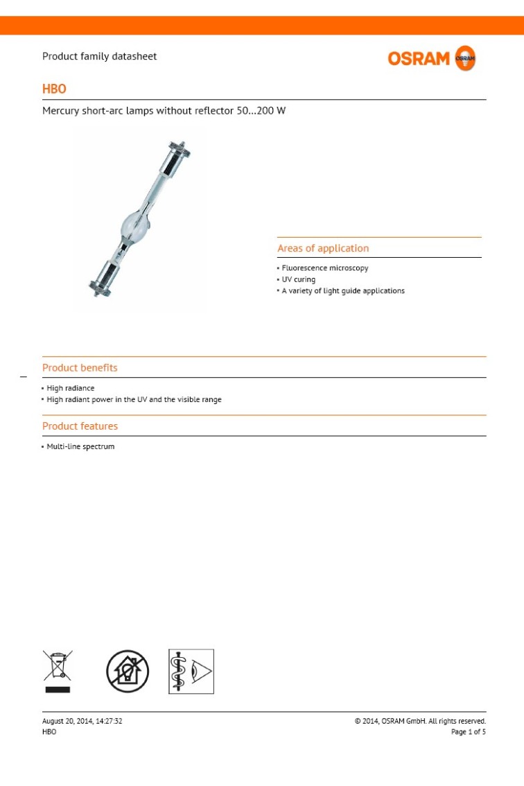
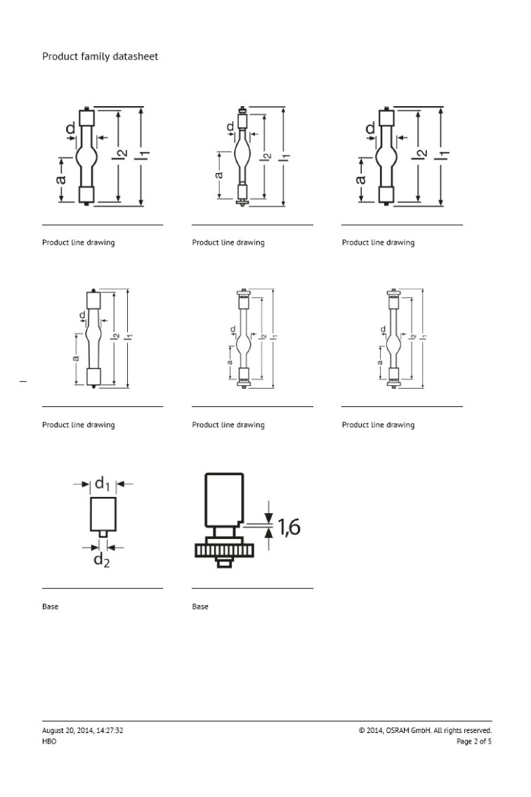
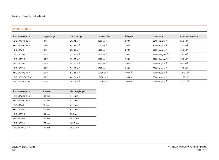
Caratteristiche delle lampade HBO OSRAM


Sub-Menu:
- Description
- Diascopy
- Epi-Fluoresc.
- Illumination ←
- Installing and Aligning a Mercury Lamp
- Fluorocromes
- Tables of Fluorocromes
- The Fading
- Wavelengths
- Properties
- % of transmission
- Types of filters
- Overlapping Spectra
- Intensity
- Image acquisition
- The Filter's set
- The Filters
- Fluorochrome's praparation
- Sample's preparations
- Fluorescence Sample
- Cytochemical Fluorescence
- Intrinsic Fluorescence
- Gallery